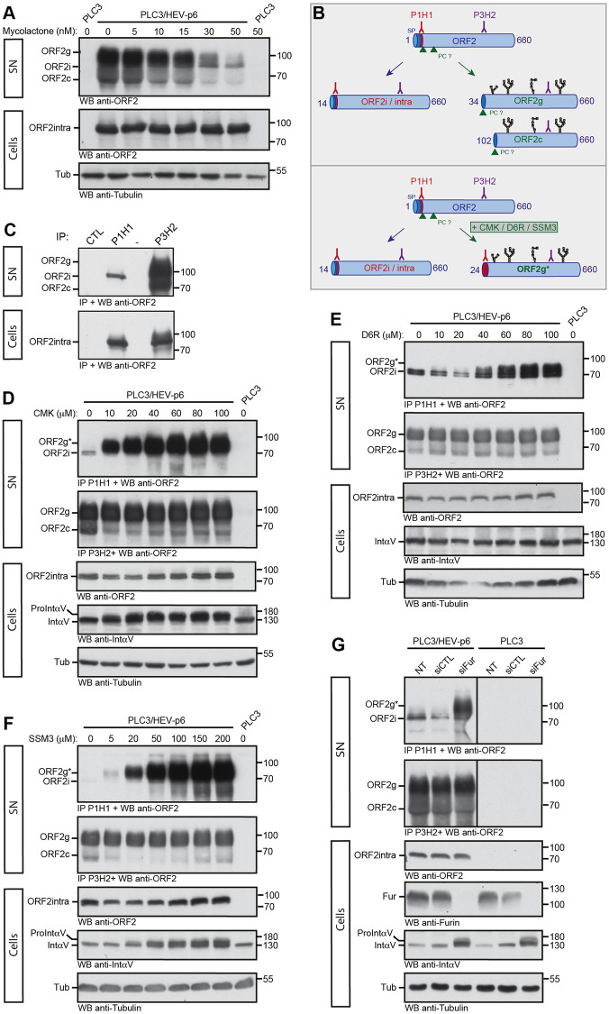
Fig 4. Translocation and maturation of the glycosylated ORF2 forms.
(A) Dose-response inhibition of ORF2g/c secretion in mycolactone-treated cells. At 7 d.p.e., PLC3/HEV-p6 and mock cells were treated for 24h with the indicated concentrations of mycolactone (in nM) or maximal volume of the vehicle, ethanol (indicated as 0 nM). Supernatants (SN) and lysates (Cells) were collected and ORF2 proteins were detected by WB using the 1E6 Ab. Tubulin served as control protein loading. (B) Schematic representation of ORF2i/g/c proteins and recognition sites of P1H1 and P3H2 antibodies used to discriminate the different ORF2 forms. SP, signal peptide. PC, proprotein convertase. Glycans are in black. (C) Immunoprecipitation of ORF2 proteins in SN and lysates of PLC3/HEV-p6 cells by P1H1, P3H2 and isotype control (CTL) antibodies immobilized on magnetic beads. ORF2 proteins were detected by WB using the 1E6 Ab. (D-F) At 7 d.p.e., PLC3/HEV-p6 cells were treated with the indicated concentrations of Decanoyl-RVKR-chloromethylketone (CMK), hexa-D-arginine amide (D6R) and SSM3 trifluoroacetate (SSM3) (in μM) or DMSO diluent (indicated as 0 μM). (G) At 7 d.p.e., PLC3/HEV-p6 and PLC3 mock cells were transfected with siRNA targeting furin (siFur) or non-targeting siRNA (siCTL) or left non-transfected (NT). (D-G) At 72h post-treatment or post-transfection, supernatants (SN) and lysates (Cells) were collected. SN were immunoprecipitated with P1H1 and P3H2 antibodies and ORF2 proteins were detected by WB using the 1E6 Ab. ORF2intra, αV-Integrin (IntαV) and Tubulin (Tub) were detected in cell lysates. In G, furin (Fur) was also detected in cell lysates. αV-pro-integrin (ProintαV) corresponds to the non-maturated αV-integrin. ORF2g* corresponds to the ORF2g immunoprecipitated by the P1H1 Ab. Molecular mass markers are indicated on the right (kDa).

