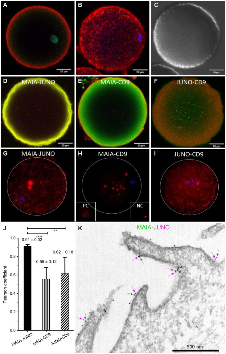
Fig. 2. MAIA immunolocalization and interaction in human oocytes.
(A to C) MAIA on human mature metaphase II (MII) stage oocyte (red), β-tubulin (green), nuclear counterstain (Hoechst 33342), confocal microscopy; (A) single plane, (B) a maximum intensity projection visualization of MAIA localization and (C) differential interference contrast (DIC). (D to F) Colocalization between protein pairs (D) MAIA-JUNO, (E) MAIA-CD9, and (F) JUNO-CD9 on human MII oocyte explained by preferential localization of the proteins in, or close to, the cell membrane (fig. S2). (G to I) PLA on human MII oocyte for protein pairs (G) MAIA-JUNO, (H) MAIA-CD9, and (I) JUNO-CD9 with appropriate negative control (NC) α-tubulin–β1 integrin and positive control (PC) α-tubulin–β-tubulin. (J) Pearson correlation coefficient for the protein pairs corresponding to colocalization analysis (fig. S2). (K) Representative transmission electron microscopy image of MAIA (big dots and green arrows) and JUNO (small dots and magenta arrows) localized on the microvilli/oolemma of MII oocyte.

