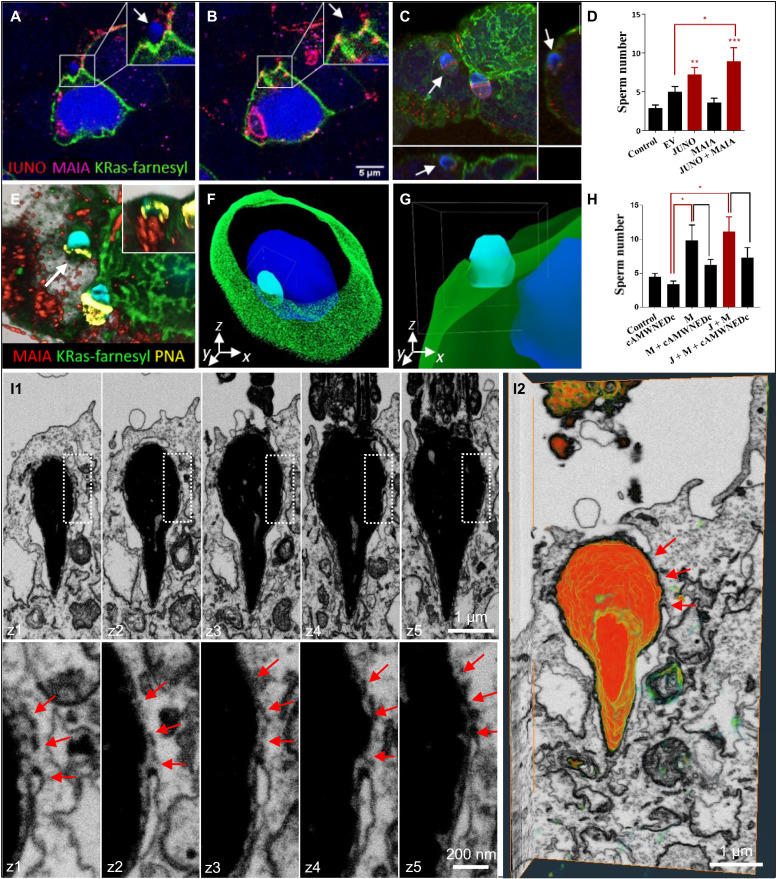
Fig. 4. Sperm binding/fusion with MAIA and JUNO cotransfected cells.
(A to C) Membrane disruption and sperm head fusion (white arrows) with cotransfected HEK293T (HEK) by MAIA (purple), JUNO (red), and cell membrane–incorporated GFP-tagged farnesylated K-Ras (green) (movie S8). (D) Quantification of sperm binding to cotransfected HEK293T (HEK) with MAIA, JUNO, and MAIA + JUNO. (E to G) Immunofluorescence data postprocessing. (E) Acrosome-reacted sperm (white arrow) equatorial segment stained with PNA (yellow) fused with cell membrane (green); the apical part of the sperm head within the cell cytoplasm is shown in the top right corner (visualized by Imaris). (F and G) Sperm-HEK penetration and fusion 3D model (Imaris), MAIA (purple), JUNO (red), and AF488-phalloidine–stained F-actin (green) (movie S9). (H) Sperm-HEK binding decrease after sperm-cAMWNEDc coincubation. (H and D) Image Xpress screening system; ImageJ/FIJI; error bars (SEM); EV, empty vector; J, JUNO; and M, MAIA. (I1 and I2) 3D visualization of the sperm head within the cytoplasm of MAIA-JUNO cotransfected HEK cell using FIB-scanning electron microscopy. (I1) Consecutive FIB-scanning electron microscopy sections showing fusion (red arrows) between the plasma membrane of MAIA-JUNO cotransfected HEK cell and the inner acrosomal membrane (IAM) of the sperm head around the equatorial segment. Overview of an internalized sperm head (top) and detail of the membrane fusion (bottom) demarcated by dotted rectangle (above). Spacing between frames, 250 nm. (I2) 3D volume rendering of the whole FIB-scanning electron microscopy dataset (x-y-z projection side view) showing partial exposure of the sperm head to the cytoplasm of the HEK cell and the site of membrane fusion (red arrows) (movie S10).

