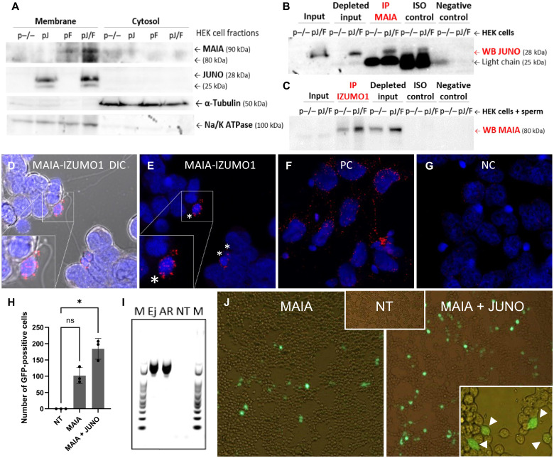
Fig. 5. MAIA-IZUMO1 protein-protein interaction and sperm-cell GFP fusion assay.
(A) Recombinant MAIA and JUNO protein localization in HEK plasmatic membrane; both proteins are shown with two isoforms (two bands) because of protein glycosylation; α-tubulin was used as a loading control for cytosolic fraction; Na/K adenosine triphosphatase (ATPase) was used as a reference protein for membrane fraction. (B and C) Co-IP of (B) MAIA-JUNO and (C) MAIA-IZUMO1 using cotransfected HEK (B) without or (C) with sperm; ISO control, isotype IgG control antibody (IgG light chain, 25 kDa; IgG heavy chain, 50 kDa). (D to G) PLA assay for MAIA-IZUMO1 on JUNO + MAIA cotransfected HEK with sperm; the positive signal is designated exclusively to the point of sperm head–cell attachment (white asterisks) (fig. S5). (D and E) MAIA-IZUMO1; (F) positive control: α-tubulin–β-tubulin; (G) negative control: MAIA–β-tubulin. (H to J) GFP fusion assay of sperm with HEK cells. (H) Number of GFP-positive cells (n) per well, nontransfected (NT), MAIA, and MAIA + JUNO cotransfected HEK cells with fused GFP-transfected sperm; *P ≤ 0.05. (I) Polymerase chain reaction (PCR) showing GFP in transfected ejaculated (Ej) and acrosome-reacted (AR) sperm, nontransfected, and marker (M). (J) Representative fluorescent images showing analyzed groups (H and I), positively transfected cells (white arrows) fused with sperm with visible tail are shown in the bottom right corner, and fluorescent signal is merged with bright field. ns, not significant.

