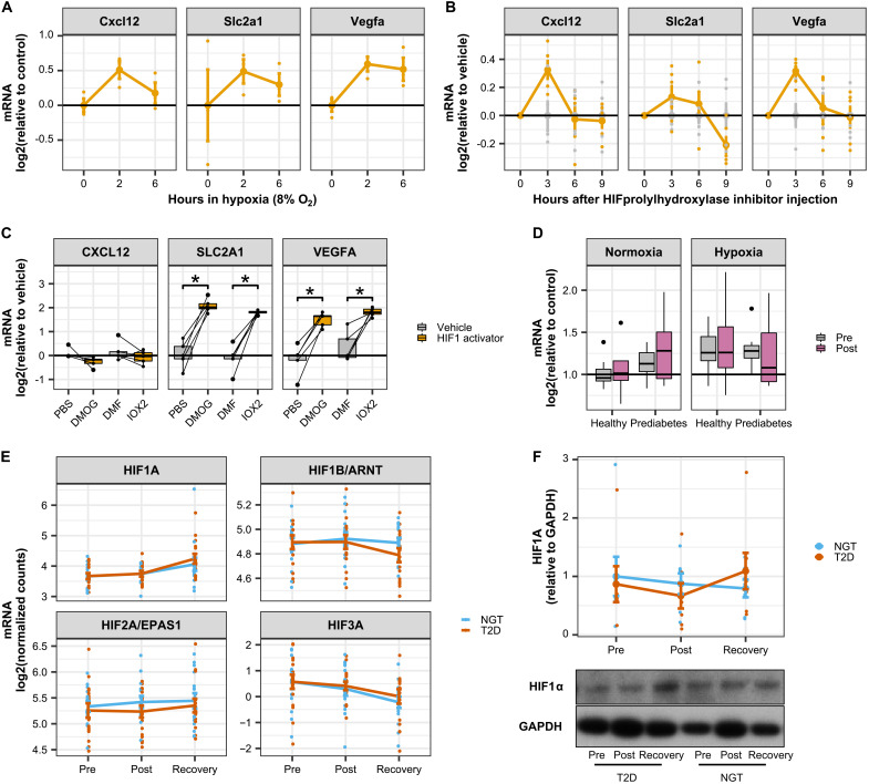
Fig. 5. CXCL12 is induced by hypoxia in skeletal muscle tissue but not in primary myotube cultures.
(A) Publicly available data (GSE81286) for cytokine mRNA expression in skeletal muscle from mice exposed to hypoxia (8% oxygen for up to 6 hours). Data are means ± SE. (B) Publicly available data (GSE95244) for cytokine mRNA expression in skeletal muscle from mice in which HIF1α was activated by an injection of an HIF prolyl hydroxylase inhibitor. Data are area under the curve of the time courses presented in fig. S3C. *P < 0.05 at least one time point after injection of the prolyl hydroxylase inhibitor. (C) Primary human skeletal muscle cells treated for 4 hours with the HIF1A stabilizers DMOG (100 μM) or IOX2 (200 μM). Box-and-whisper plots from five independent donors. *P < 0.05 paired t test versus respective vehicles. (D) CXCL12 mRNA expression in human skeletal muscle. Biopsies were collected before and after an acute bout of exercise in men with prediabetes versus metabolically healthy controls. Half of the participants performed exercise under hypoxia. Box-and-whisper plots from seven to eight independent donors. (E) HIF mRNA in skeletal muscle biopsies from men with type 2 diabetes (n = 20) versus healthy individuals (n = 18) from RNA sequencing data presented in Fig. 2. Data are means ± SE. (F) HIF1A protein abundance in skeletal muscle biopsies from men with type 2 diabetes (n = 7) versus healthy individuals (n = 7). Data are means ± SE.

