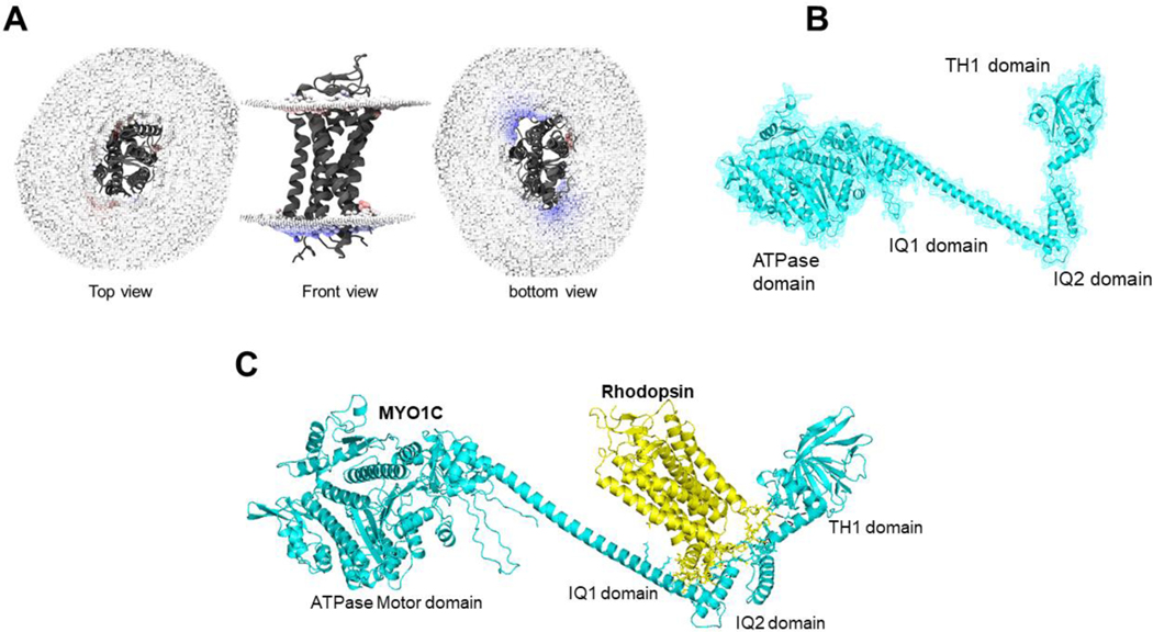
Figure 4: Homology modeling of MYO1C and Rhodopsin and their predicted C-terminus interaction.
(A) The bovine Rhodopsin structure in lipid bilayer and possible sites of anchoring are shown in distortion view from http://memprotmd.bioch.ox.ac.uk (146). (B) The predicted structure co-ordinates of MYO1C was obtained from the PDB database. (C) Docking of MYO1C to Rhodopsin. The complete structure of both MYO1C and Rhodopsin was obtained from https://alphafold.ebi.ac.uk/ (144).

