FIGURE 2.

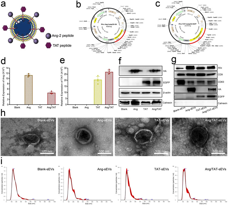
Characterisation of engineered HEK293T cells and peptide‐modified sEVs. (a, b, c) The schematic of Ang/TAT‐sEVs, the plasmid of pLVX‐IRES‐Ang‐Lamp2b‐HA, and the plasmid of pLVX‐IRES‐TAT‐Lamp2b‐EGFP. (d, e) The mRNA expression level of the Ang‐Lamp2b‐HA and TAT‐Lamp2b‐EGFP in HEK293T cells (n = 4). (f) HA and EGFP‐tag protein expression level in HEK293T cells, with β‐actin as the normalisation control. (g) HA and EGFP Tag protein and small extracellular vesicles biomarker such as CD63, CD9, Alix and negative marker (calnexin) were assessed by western blot. (h) The morphology of sEVs detected with TEM. Scale bar = 100 nm. (i) Size distribution of blank‐sEVs, Ang‐sEVs, TAT‐sEVs, and Ang/TAT‐sEVs detected through NTA
