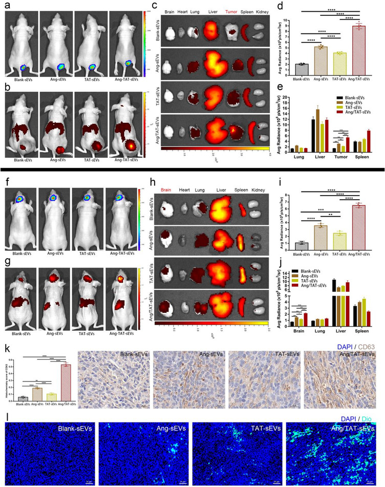
FIGURE 6.

Tumour‐targeting and BBB‐permeating ability of the peptide‐modified sEVs in vivo. (a) In vivo imaging of subcutaneous U87MG glioma identified by injecting luciferase substrate D‐luciferin potassium. (b) In vivo imaging of DiR‐labelled sEVs. (c) Ex vivo fluorescence imaging of organs and tumours from the model mice after DiR‐labelled sEVs intravenous injection. (d) Quantified data of fluorescence signal in (b) (n = 6, ****P < 0.0001). (e) Quantified data of fluorescence signal in (c) (n = 6, **P < 0.01, ***P < 0.001, ****P < 0.0001). (f) In vivo imaging of orthotopic U87MG glioma identified by injecting the luciferase substrate D‐luciferin potassium. (g) In vivo imaging of the DiR‐labelled sEVs. (h) Ex vivo fluorescence imaging of organs and tumour from the model mice after DiR‐labelled sEVs intravenous injection. (i) Quantified data of fluorescence signal in (g) (n = 4, **P < 0.01, ***P < 0.001, ****P < 0.0001). (j) Quantified data of fluorescence signal in (h) (n = 4, *P < 0.05, ***P < 0.001, ****P < 0.0001). (k) Immunohistochemistry images of CD63 expression in the brain tumour (n = 4, **P < 0.01, ****P < 0.0001). (l) Fluorescence images of DiO‐labelled sEVs targeting and penetrating the brain tumour observed through GLSM
