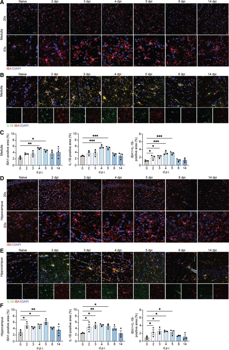
Figure 2.
Microglia contribute to neuroinflammation in the medulla oblongata and hippocamus of SARS-CoV-2-infected hamsters. Representative images of IBA1 in the hamster inferior olivary nuclei (ION) (A) and hippocampus (D) at naïve, 2, 3, 4, 5, 8 and 14 dpi, showing staining for IBA1 (red) and DAPI (blue) at ×20 and ×63. Immunostaining for IL-1β and IBA1 in the hamster ION (B) and hippocampi (E) at naïve, 2, 3, 4, 5, 8 and 14 dpi, presented as microscopy with IBA1 (red), IL-1β (green) and DAPI (blue). Quantitation of per cent IBA1+ and IL-1β+ areas, and IL-1β+IBA1+ area, normalized to total IL-1B+ area for ION (C) and hamster (F). Data were pooled from at least two independent experiments. Scale bars = 20 μm (×20) or 10 μm (×63). Data represent the mean ± SEM and were analysed by two-way ANOVA or Student’s t-test.

