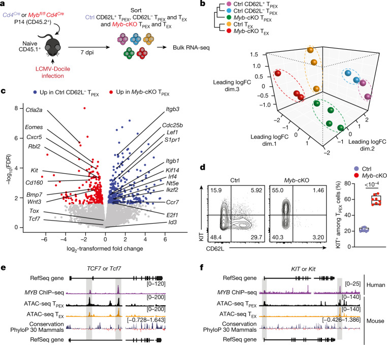
Fig. 3. MYB regulates the expression of genes that are critical for the function and maintenance of exhausted T cells.
a–c, Congenically marked Mybfl/flCd4Cre (Myb-cKO) and Cd4Cre (control) P14 T cells were adoptively transferred into naive recipient mice, which were then infected with LCMV-Docile. Splenic P14 subsets were sorted at 7 dpi and processed for bulk RNA-seq. a, Schematic of the experimental set-up. b, Sample dendrogram and three-dimensional scaling plot of all the samples. logFC, log-transformed fold change. c, Volcano plot highlighting genes that are differentially expressed (false discovery rate (FDR) < 0.15) between Myb-cKO TPEX and control CD62L− TPEX cells, with genes of interest annotated. d, Flow cytometry plots and quantification show the frequencies of KIT+ cells among control and Myb-cKO TPEX P14 T cells at day 8 after infection with LCMV-Docile (gated on TPEX cells). e,f, Tracks show MYB chromatin immunoprecipitation followed by sequencing (ChIP–seq) peaks in the TCF7 (e) and KIT (f) gene loci of human Jurkat T cells and assay for transposase-accessible chromatin using sequencing (ATAC-seq) peaks of TPEX and TEX cells in the corresponding mouse gene loci aligned according to sequence conservation. Dots in graph represent individual mice; box plots indicate minimum and maximum values (whiskers), interquartile range (box limits) and median (centre line). Data are representative of two independent experiments (d). P values are from two-tailed unpaired t-tests (d).

