FIGURE 1.

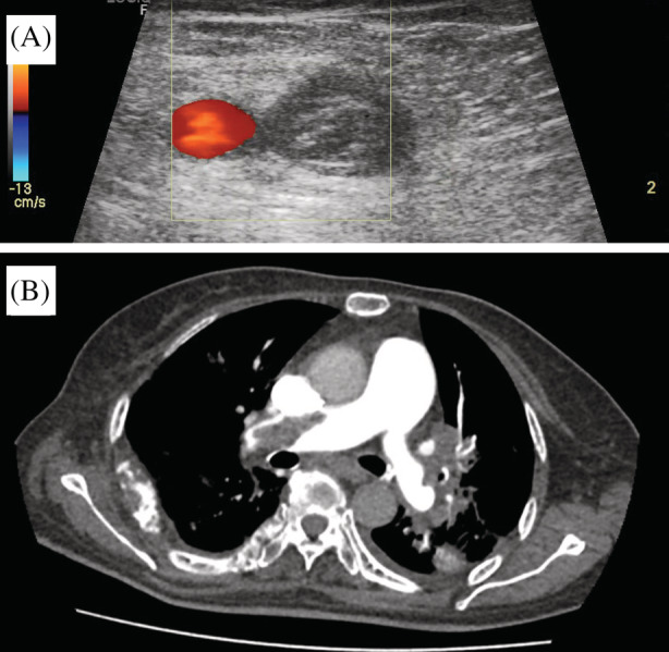
(A) Lower limb POCUS confirmed left lower limb DVT with direct visualization of a thrombus in the left femoral vein. (B) Representative slice of the patient's CTPA (axial view) confirmed a large thrombus in the right main pulmonary artery

(A) Lower limb POCUS confirmed left lower limb DVT with direct visualization of a thrombus in the left femoral vein. (B) Representative slice of the patient's CTPA (axial view) confirmed a large thrombus in the right main pulmonary artery