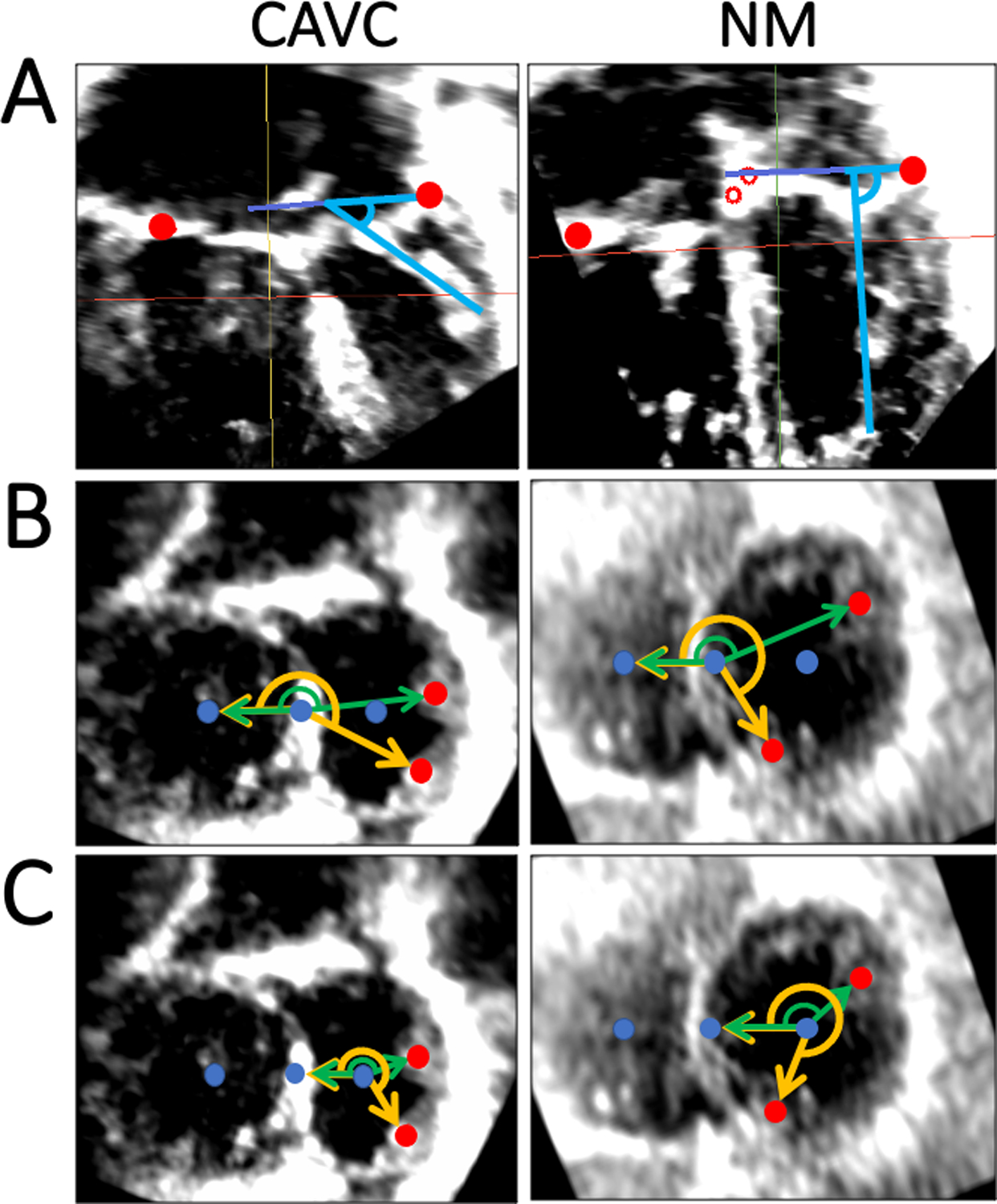
Figure 4. Papillary Muscle Angle Quantification in CAVC and Normal Mitral Valves.

A. Papillary muscle angle relative to annulus plane in CAVC and normal mitral; B. “Septal Point” based rotation angle measurement in CAVC and normal mitral valves. Reference vector is perpendicular to the septal plane, and papillary muscle base points are projected to the common CAVC and common normal mitral/tricuspid valve plane; C. “Center point” based rotation angle measurement in CAVC and normal mitral valves. Reference vector is perpendicular to the center plane, and papillary muscle base points are projected to the left CAVC and mitral valve plane. CAVC = Complete atrioventricular canal, NM = Normal Mitral.
