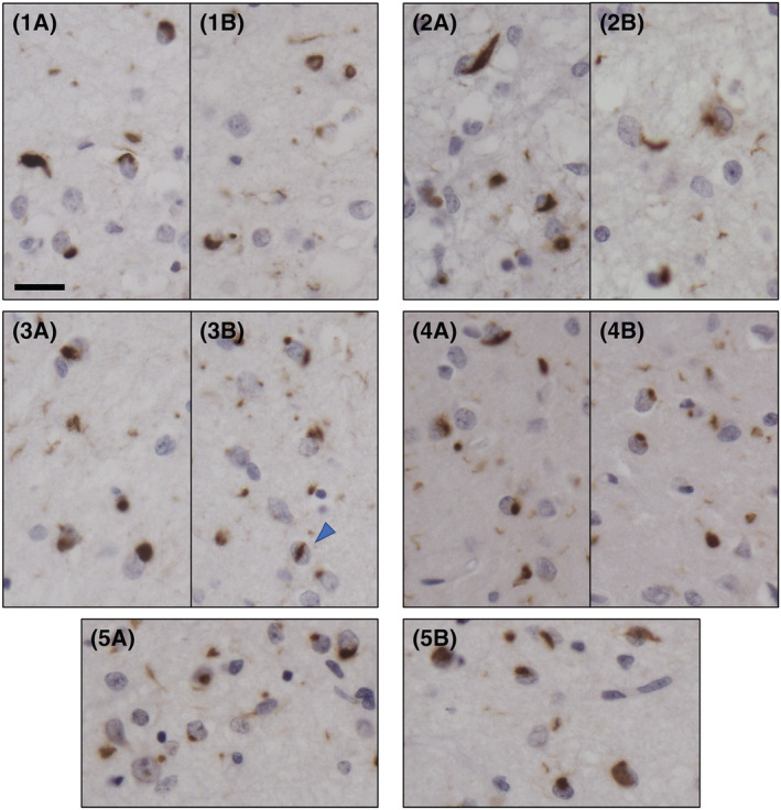
FIGURE 2.

TDP‐43 immunohistochemistry in the frontal cortex of five GRN mutation carriers (Cases 1–5). TDP‐43 immunoreactive intracellular inclusions and neuropil deposits in two fields of the second layer of the frontal cortex (A, B). Neuronal cytoplasmic inclusions (NCIs), a neuronal intranuclear inclusion (NII, arrowhead) and granular deposits are seen in the neuropil. The pattern of TDP‐43 inclusions is consistent with Mackenzie Type A classification. Scale = 20 μm; antibody: pS409/410
