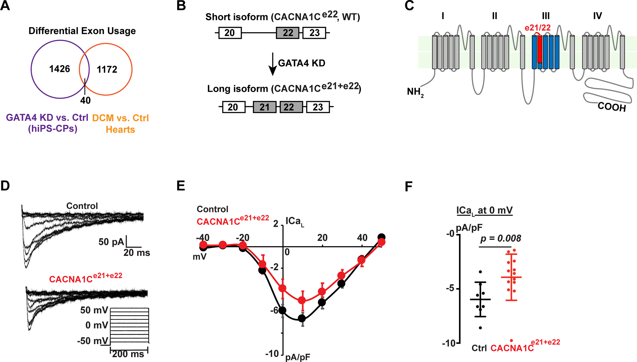
Fig. 5: GATA4 regulated physiologically-relevant alternative splicing, resulting in functionally distinct protein products.

(A) Intersection of alternatively spliced exons upon GATA4-knockdown (KD) and alternatively spliced exons between human dilated cardiomyopathy (DCM) patient hearts and healthy donor hearts. (B) Schematic of short (WT) and long (CACNA1C e21+e22) isoforms of CACNA1C with differential inclusion of exon 21. (C) Predicted protein topology of the CACNA1C, showing the exon21/22 (red) localizing to the IIIS2 transmembrane segment and part of the linker region between IIIS1 and IIIS2. (D) Whole cell calcium current recordings from neonatal rat ventricular myocytes infected with adenovirus containing green fluorescent protein (GFP) or GFP + CACNA1Ce21+e22. Voltage protocol used to elicit L-type calcium current is shown at the bottom, right. (E) Current-voltage relationships for GFP infected (black, n=8) or CACNA1Ce21+e22 infected (red, n=13) neonatal rat ventricular myocytes (NRVMs), normalized to cell capacitance. Data are shown as means ± SEM. (F) Peak L-type calcium current density at 0 mV for GFP infected (black, n=8) or CACNA1Ce21+e22 infected (red, n=13) NRVMs. Error bars denote standard deviation. P=0.008 by Mann-Whitney test.
