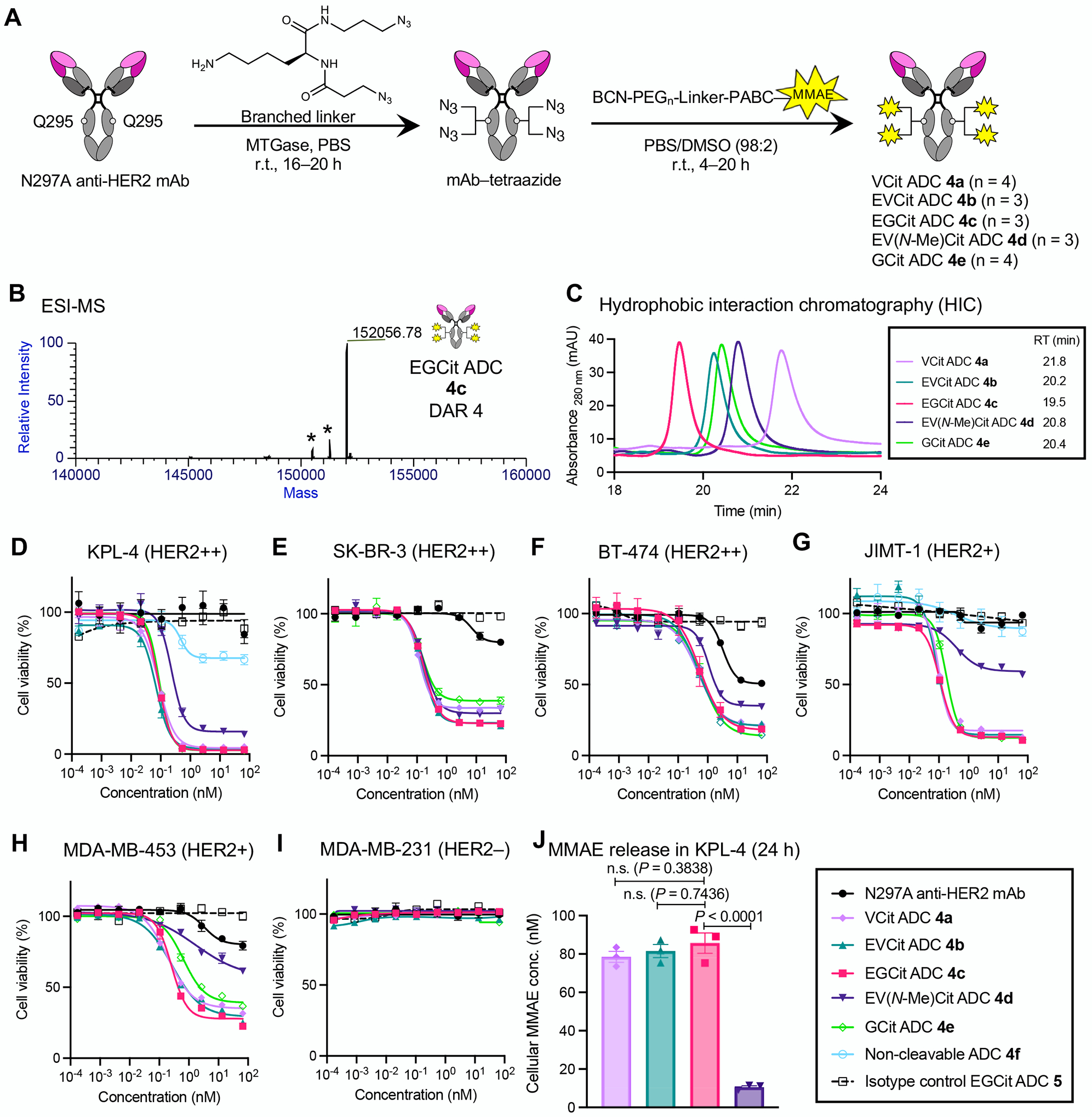
Fig. 3.

EGCit linker increases ADC hydrophilicity and cell killing potency with efficient intracellular payload release. A Construction of ADCs (4a–e) by MTGase-mediated branched linker conjugation and following strain-promoted azide–alkyne cycloaddition (yellow spark: MMAE). B Deconvoluted ESI-MS trace of EGCit ADC 4c. Asterisk (*) indicates a fragment ion detected in ESI-MS analysis. See Supplementary Notes for mass traces of the other ADCs. C Overlay of five HIC traces (VCit ADC 4a: light purple; EVCit ADC 4b: green; EGCit ADC 4c: magenta; EV(N-Me)Cit ADC 4d: purple; GCit ADC 4e: light green) under physiological conditions (phosphate buffer, pH 7.4). D–I Cell killing potency in the breast cancer cell lines KPL-4 (D), SK-BR-3 (E), BT-474 (F), JIMT-1 (G), MDA-MB-453 (H), and MDA-MB-231 (I). We tested unconjugated N297A anti-HER2 mAb (black circle), VCit ADC 4a (light purple diamond), EVCit ADC 4b (green triangle), EGCit ADC 4c (magenta square), EV(N-Me)Cit 4d (purple inversed triangle), GCit ADC 4e (light green open diamond), non-cleavable ADC 4f (cyan open circle), and isotype control EGCit ADC 5 (black open rectangle with dotted curve). J ESI-MS-based quantification of free MMAE released from ADCs 4a–c in KPL-4 cells after incubation at 37 °C for 24 h. All assays were performed in triplicate. Data are presented as mean values ± SEM. For statistical analysis, a one-way ANOVA with a Dunnett’s post hoc test was used (comparison control: EGCit ADC 4c). BCN, bicyclo[6.1.0]nonyne; DAR, drug-to-antibody ratio; MMAE, monomethyl auristatin E; MTGase, microbial transglutaminase; PEG, polyethylene glycol; RT, retention time.
