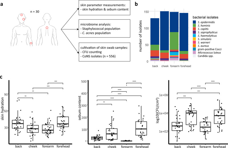
Fig. 1. Skin parameters and distribution of staphylococcal isolates on four different skin sites.
a Study design. b Number of isolates of identified bacterial species per skin site. A selective cultivation approach to primarily isolate staphylococci was applied. c Skin hydration, sebum content and CFU per cm2 on back, cheek, forearm, and forehead skin (n = 30 for each skin site. *p ≤ 0.05, **p ≤ 0.01, ***p ≤ 0.001, ****p ≤ 0.0001. Unpaired Wilcoxon test). Middle lines of boxplots indicate the median. Lower and upper lines represent the first and third quartiles. Whiskers show the 1.5x inter-quartile ranges.

