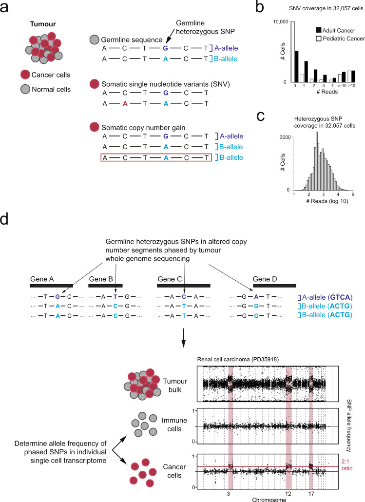
Fig. 1. Overview of different approaches to identifying cancer-derived cells.
a Genomic changes present in cancer genomes. b Number of cells (y-axis) with N reads covering point mutations (x-axis), separated by low (NB neuroblastoma) and high (RCC renal cell carcinoma) mutation burden. c Number of cells (y-axis) with N reads covering heterozygous single nucleotide polymorphisms (SNP) (x-axis). d Overview of using allelic shifts representing copy-number changes to detect cancer cells.

