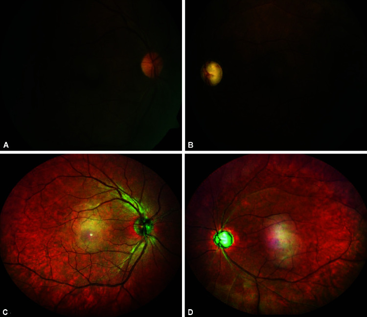
Figs 1A to D.
(A) Clinical photograph showing normal disk in the right eye; (B) Glaucomatous disk in the left eye with attenuated arterioles; (D) Note the altered reflective signals from the ischemic retina (arrow) in the left eye; (C and D) Central ghost maculopathy artifacts (arrowhead) seen on wide-field multicolor imaging in both eyes

