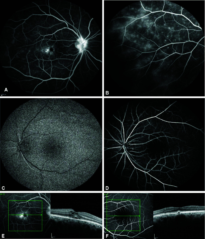
Figs 2A to F.
(A) Fundus fluorescein angiography (FFA) shows disk leakage, perifoveal leakage; and (B) microvascular leakages in the right eye; and (C) delayed arm retinal; and (D) prolonged arteriovenous transit time in the left eye (E) optical coherence tomography (OCT) shows cystoid macular edema (CME) with submacular detachment in the right eye; and (F) inner retinal minimal reflectivity and atrophy with small pigment epithelial detachment in the left eye

