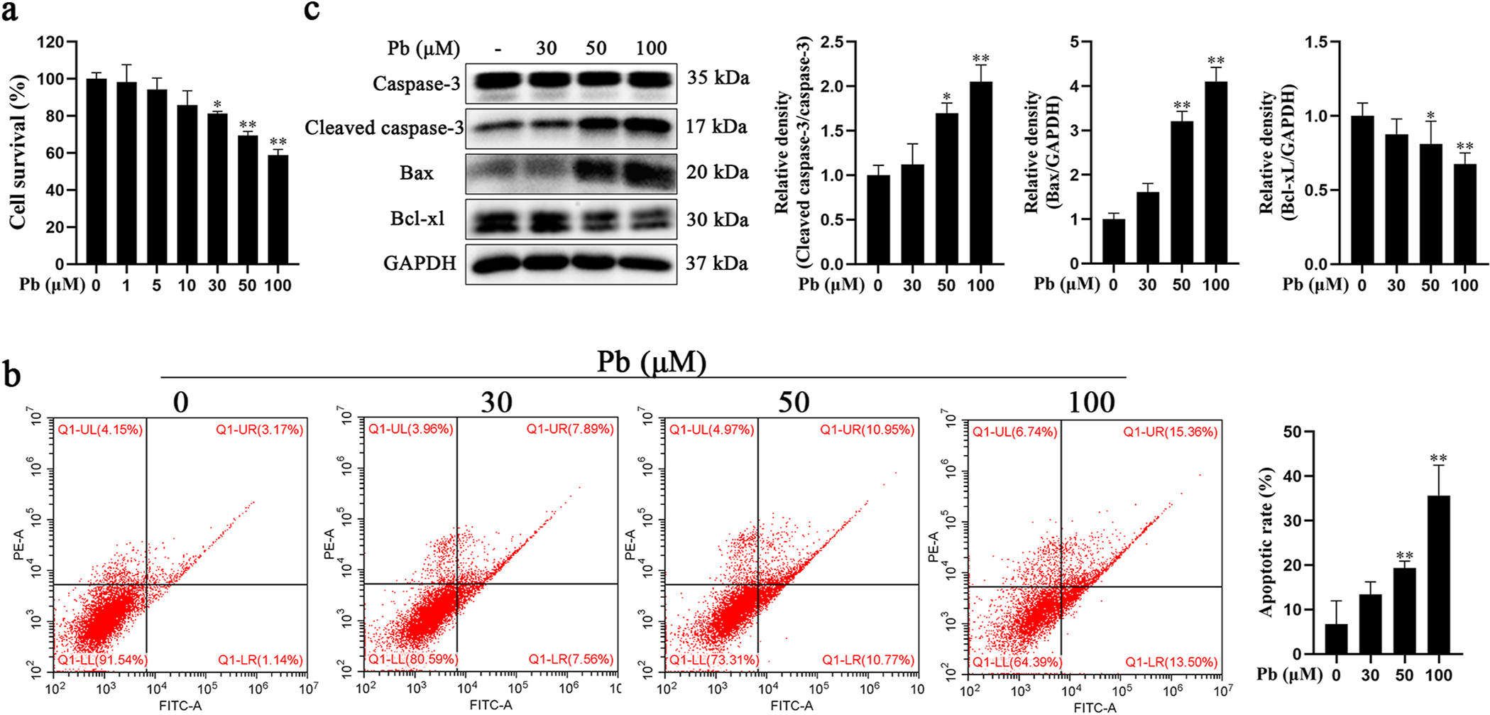
Fig. 2.

Pb exposure induced apoptosis in primary hippocampal neurons. a-c Primary hippocampal neurons were treated with increased concentrations of PbAc for 24 h. a Cell viability was determined with CCK8. b Neuronal apoptotic rate was determined by flow cytometry analysis. Early apoptotic cells (AV+/PI ) are shown in the lower right quadrant and late apoptotic cells (AV+/PI+) in upper right quadrant. c Protein levels of cleaved caspase-3, Bax and Bcl-xL were determined by western blotting. The protein expression was normalized by GAPDH or corresponding total protein content. * P < 0.05 and * * P < 0.01, compared to the control group.
