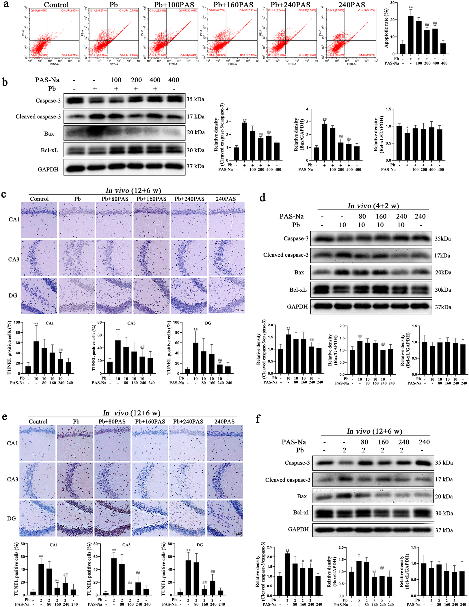
Fig. 3.

PAS-Na treatment decreased the Pb-induced hippocampal neuron apoptosis. a, b Primary hippocampal neurons were treated with 50 μM PbAc for 24 h, followed by 100, 200 or 400 μM PAS-Na treatment for 24 h. a Apoptosis in hippocampal neurons was determined using flow cytometry analysis. b Protein levels of cleaved caspase-3, Bax and Bcl-xL were determined by western blotting. c, d Rats were treated with 10 mg/kg PbAc for 4 weeks during juvenile, followed by 80, 160 or 240 mg/kg PAS-Na treatment for 2 weeks. e, f Rats were treated with 2 mg/kg PbAc for 12 weeks, followed by 80, 160 or 240 mg/kg PAS-Na treatment for 6 weeks. c, e Apoptotic nuclei in hippocampal tissues of rats were determined using TUNEL assay. Brown-red-stained cell nuclei were considered positive apoptosis cells, while blue-stained nuclei were identified as negative cells. Scale bar = 50 μm. d, f Protein level of cleaved caspase-3, Bax and Bcl-xL were determined by western blotting. The protein expression was normalized by GAPDH or corresponding total protein content. * P < 0.05 and * * P < 0.01, compared to the control group. #P < 0.05 and ##P < 0.01, compared to the Pb-treated group.
