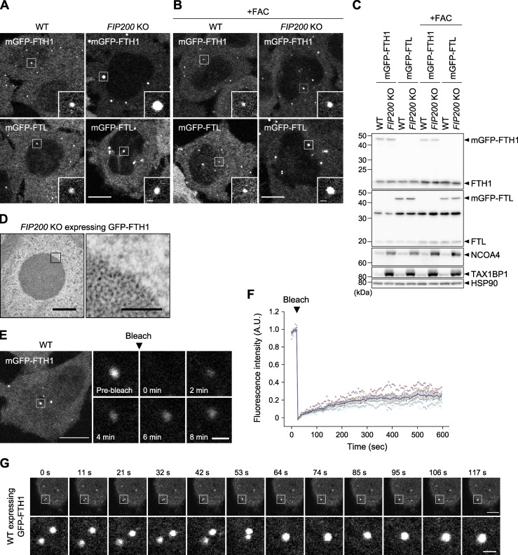
Figure 1.
Ferritin particles assemble to form liquid-like condensates. (A and B) Fluorescent images of the ferritin subunits FTH1 and FTL. WT and macroautophagy-deficient FIP200 KO HeLa cells stably expressing mGFP-FTH1 or mGFP-FTL were grown in DMEM (A) or treated with 10 μg/ml ferric ammonium citrate (FAC) for 24 h (B) and observed by fluorescence microscopy. Scale bars, 10 μm (main) and 1 μm (inset). (C) Immunoblots showing the expression levels of mGFP-FTH1 and mGFP-FTL in cells used in A. (D) TEM images of the ferritin condensates in FIP200 KO HeLa cells expressing GFP-FTH1. Scale bars, 500 nm (main) and 100 nm (magnified). (E) FRAP analyses of the ferritin condensates. WT HeLa cells expressing mGFP-FTH1 were treated with 100 μg/ml FAC for 24 h followed by 50 μM deferoxamine (DFO) for 5 h, and subjected to FRAP analyses. Scale bars, 10 μm (main) and 1 μm (magnified). (F) Quantification of E. Fluorescence intensities before photobleaching were set to 1. Representative results from two independent experiments are presented as means ± SEM (n = 5). (G) Coalescence of the ferritin condensates. WT HeLa cells expressing GFP-FTH1 were grown in DMEM and observed by time-lapse fluorescence microscopy at ∼11-s intervals. Scale bars, 10 μm (main) and 1.5 μm (magnified). Source data are available for this figure: SourceData F1.

