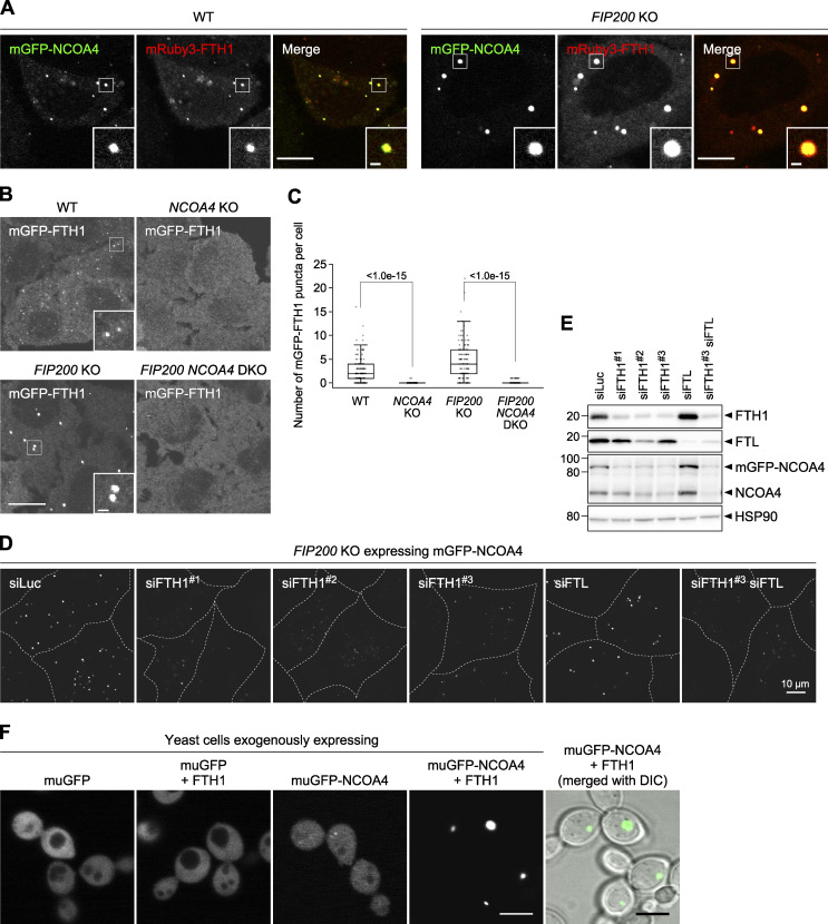
Figure 2.
NCOA4 drives ferritin condensate formation. (A) Localization of NCOA4 in ferritin condensates. WT and FIP200 KO HeLa cells expressing both mGFP-NCOA4 and mRuby3-FTH1 were grown in DMEM and observed by fluorescence microscopy. Scale bars, 10 μm (main) and 1 μm (inset). (B) NCOA4 is essential for condensate formation. WT, NCOA4 KO, FIP200 KO, and FIP200 NCOA4 double KO (DKO) HeLa cells expressing mGFP-FTH1 were observed as in A. Scale bars, 15 μm (main) and 1.5 μm (inset). (C) The number of mGFP-FTH1 puncta per cell in B was quantified (n = 112–210 cells from three biological replicates). Solid bars indicate the means, boxes the interquartile ranges, and whiskers the 10th to 90th percentiles. Differences of puncta counts among the cells were statistically analyzed by Welch’s t test with Holm–Sidak method for multiple comparison (two-tailed test). Data distribution was assumed to be normal, which was not formally tested. (D) Knockdown of ferritin subunits. FIP200 KO HeLa cells expressing mGFP-NCOA4 were transfected with the indicated siRNAs (siLuc, siFTH1 [three independent oligos were used], and siFTL) and observed by fluorescence microscopy. Scale bar, 10 μm. (E) Immunoblots showing the expression levels of FTH1, FTL, and NCOA4 in cells used in D. (F) NCOA4 and FTH1 are sufficient for condensate formation. Yeast cells exogenously expressing muGFP-NCOA4 with or without FTH1 were observed by fluorescence microscopy. Scale bar, 5 μm. Source data are available for this figure: SourceData F2.

