Figure S1.
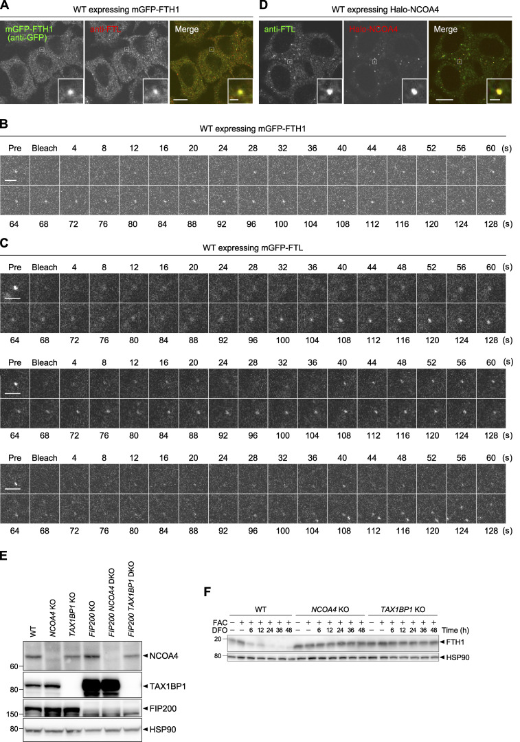
Ferritin forms liquid-like condensates with NCOA4. (A and D) WT HeLa cells expressing mGFP-FTH1 (A) or Halo-NCOA4 (D) were grown in DMEM, treated with 100 nM HaloTag SaraFluor 650T ligand (D), and then subjected to immunostaining with anti-FTL monoclonal antibody. Scale bars, 10 μm (main) and 1 μm (inset). (B and C) FRAP analyses of ferritin condensates. WT HeLa cells expressing mGFP-FTH1 (B) or mGFP-FTL (C) were treated with 100 μg/ml FAC for 24 h followed by 50 μM DFO for 2 h, and subjected to FRAP analyses (4-s intervals for 128 s). Three individual mGFP-FTL puncta are shown in C. Scale bars, 2 μm. (E) WT, NCOA4 KO, TAX1BP1 KO, FIP200 KO, FIP200 NCOA4 DKO, and FIP200 TAX1BP1 DKO HeLa cells were grown in DMEM. Whole-cell lysates were analyzed by immunoblotting with antibodies against NCOA4, TAX1BP1, FIP200, and HSP90. (F) WT, NCOA4 KO, and TAX1BP1 KO cells were grown in DMEM and treated with 10 μg/ml FAC for 24 h followed by 50 μM DFO for the indicated hours. Whole-cell lysates were analyzed by immunoblotting with antibodies against FTH1 and HSP90. Source data are available for this figure: SourceData FS1.

