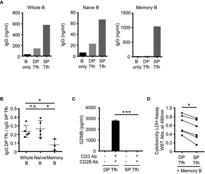
Figure 4.
Functional effects of DP-Tfh cells on B-cell regulation. (A) Representative graphs of co-culture experiments to examine antibody production using autologous Tfh and B cells sorted from tonsil specimens (n = 3-5). DP-Tfh cells (CD3+CD4+CD8+CXCR5hiPD-1hi) or SP-Tfh cells CD3+CD4+CD8-CXCR5hiPD-1hi) were co-cultured with whole B cells (CD3-CD19+), naïve B cells (CD3-CD19+IgD+CD27-), or memory B cells (CD3-CD19+IgD-CD27+) under the stimulation of CD3, CD28, and CD40L. After incubating cells for 7 days, IgG levels in the supernatants were analyzed by ELISA. (B) Effects of DP-Tfh cells on B cells investigated by co-culture experiments as demonstrated in (A). Data indicate ratios of IgG levels from whole B cells, naïve B cells, or memory B cells in the presence of DP-Tfh cells to those in the presence of SP-Tfh cells (IgG DP-Tfh/IgG SP-Tfh). Data were obtained from three to five independent experiments using autologous tonsillar lymphocytes. (C) Increased capacity of DP-Tfh cells to secrete granzyme B (GZMB) under CD3 and CD28 stimulation in comparison with SP-Tfh cells of tonsils. After incubating cells for 7 days, GZMB levels in culture supernatants were analyzed by ELISA. Data were obtained from four independent experiments using autologous tonsillar lymphocytes. (D) Cytotoxicity of DP-Tfh cells for memory B cells in comparison with SP-Tfh cells. Co-culture supernatants of memory B cells and DP-Tfh cells or SP-Tfh cells derived from autologous tonsillar lymphocytes were analyzed by a cytotoxicity LDH/WST assay. The absorbance values indicating the cytotoxic activities of DP-Tfh and SP-Tfh cells in each experiment (depicted as a closed circle and rectangle, respectively) are connected by a line to evaluate their differences. Data were obtained from six independent experiments. Statistical significance in (B–D) was determined by the Mann–Whitney U test.

