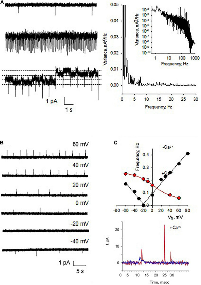
FIGURE 4.
Spontaneous electric oscillations of reconstituted DS. (A, Left) Current oscillations (in voltage-clamp mode) in the form of spikes were observed in the reconstituted membranes (n = 36), which were independent of ion channel recordings, as observed in the bottom tracing where two channel levels were indicated by the horizontal lines. Right. Power spectra of tracings at Left, disclosing spontaneous oscillations with peak frequency at 1-2 Hz, and ion channel activity (Inset, showing a Lorentzian shoulder). (B) The spontaneous electrical oscillations were detected in the absence of NMDA added from the trans side of the chamber, whose frequency depended on the holding potential. (C) Spontaneous activity was strongly dependent on the presence of external (trans) Ca2+ (15 mM). The amplitude (as well as polarity) and frequency were strongly dependent on the presence of Ca2+. (Top panel) The black solid lines were the best linear fitting of the recorded frequencies vs. holding potential, with the function –0.0042V – 0.0749 (R = 0.9867) for negative values, and 0.0041V + 0.0949 (R = 0.9935) for positive values, respectively. The red line was the best fitting with a sigmoid function 0.0471 + 0.2150 (1 + exp(–3.6039V/21.1850))– 1 (R = 0.9994). Bottom panel shows representative Ca2+-dependent frequency spikes in the absence (Blue), and presence (Red) of the ion.

