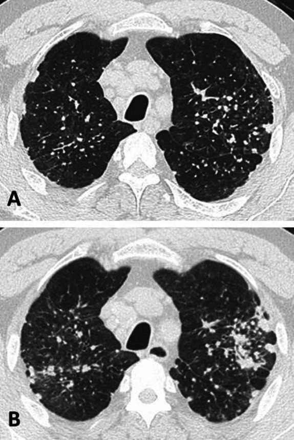
Figure 1.

High-resolution chest CT image (A) from an engineered stone worker shows small rounded opacities in a perilymphatic distribution, interlobular septal thickening, mediastinal lymphadenopathy and areas of emphysema. Repeat chest CT scan 3 years later (B) shows increased ground glass nodularity and progression of bilateral conglomerate opacities despite removal from workplace exposure to respirable crystalline silica.
