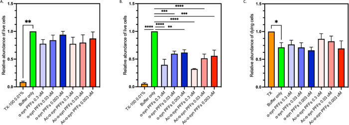
Figure 8.
N-terminal acetylation does not modify the cytotoxicity of α-synuclein fibrils and fibril fragments. (A, B) Relative end-point fluorescence intensity of Calcein AM and a live cell dye expressed in arbitrary fluorescence units, in the presence of Triton X-100 (orange) or PBS (green) or when incubated with at 0.3–0.003 μM sonicated fibrils (PFFs) of non-acetylated α-synuclein (blue) and acetylated (Ac) α-synuclein (red) for (A) 24 h and (B) 48 h. (A) All treatments showed significantly different fluorescence levels to Triton X 100 treated cells (p < 0.05). However, a significant difference was not found between any other treatments (p > 0.05). (B) All treatments, showed significantly different fluorescence levels to buffer only control cells (p < 0.05). However, a significant difference was not found between acetylated and non-acetylated preformed sonicated fibril (PFF) treatments (p > 0.05). (C) Relative fluorescence intensity of PI expressed in arbitrary fluorescence units, a dead cell detecting dye, in the presence of Triton X-100 (orange) or PBS (green) or when incubated with at 0.3–0.003 μM sonicated fibrils (PFFs) formed from non-acetylated α-synuclein (blue) and acetylated (Ac) α-synuclein (red) for 24 h. * Indicates a p value of 0.01; other pairwise comparisons showed no significant differences (p > 0.05).

