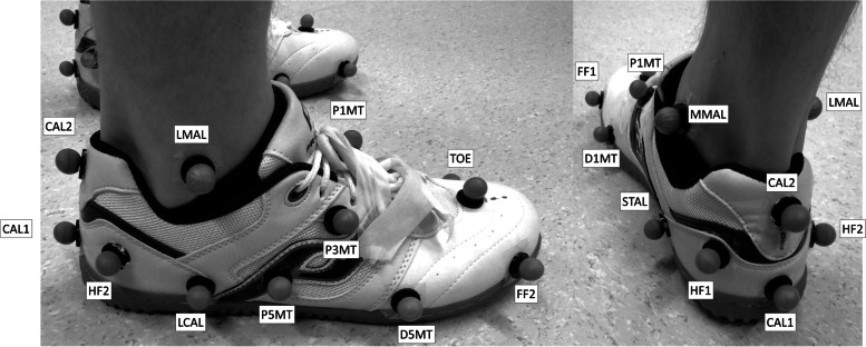
Fig. 2.
Diagram illustrating the lateral (left) and posterior-medial marker positions of the Calibrated Anatomical System Technique (CAST) multi-segment foot model and the Oxford Foot Model (OFM) applied simultaneously. CAL1 and CAL2 represents the inferior and superior posterior aspect of calcaneus; HF1 and HF2 represents the medial and lateral hindfoot; STAL represents the sustentaculum tali; LCAL represents the lateral aspect of the calcaneus (at the same distance from the most posterior point as STAL); MMAL and LMAL represents the medial and lateral malleoli; P1MT, P5MT, and P3MT represents the base of the 1st and 5th metatarsals, and between the base of the 3rd and the 4th metatarsals, respectively; D1MT and D5MT represents the medial first and lateral fifth metatarsal heads; TOE represents the mid-point of the distal heads of the 2nd and 3rd metatarsals; FF1 and FF2 were not used for the analysis

