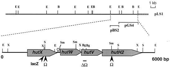
FIG. 1.
Organization of the hut locus. Restriction map of the 26.5-kb EcoRI fragment of S. meliloti Rm5000 cloned into pLAFR1 is shown above the physical and genetic organization of the hut locus. The genes deduced from the nucleotide sequence analysis are represented by boxed arrows. The positions of the Ω insertions and lacZ fusion are indicated below. The hairpin indicates the position of a stem-loop structure able to form a secondary structure. Abbreviations for restriction enzymes: Bg, BglII; E, EcoRI; S, StuI; Sm, SmaI; X, XhoI.

