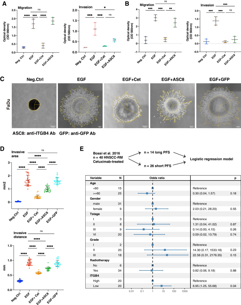
Fig. 8.
ITGB4 represents a target and potential predictive marker for HNSCC. A-B Wildtype Kyse30 and FaDu cells were analyzed in migration assays (A) and in invasion assays with inlays coated with Matrigel (B) in a Boyden chamber. Where indicated, cells were treated EGFhigh or combinations of EGFhigh with Cetuximab or anti-ITGB4 antibody ASC8. Mean and SD are shown in scatter dot plots of n = 3 independent experiments. * ≤ 0.05, ** ≤ 0.01; *** ≤ 0.001, **** ≤ 0.0001. C Spheroids of wildtype FaDu cells were cultured in Matrigel under serum-free conditions. Spheroids were left untreated (Neg.Ctrl), treated with EGFhigh, or treated with a combination of EGFhigh and Cetuximab, ASC8, or anti-GFP antibody. Representative images of invasive cells upon treatment (n = 3 independent experiments with multiple spheroids) are shown. D Invasive area representing the outer rim of cells (see yellow lines in C) and invasive distance representing the mean distance covered by 10–15 most invasive single cells were quantified. Mean and SD are shown in scatter dot plots of n = 3 independent experiments where each dot represents one spheroid. **** ≤ 0.0001. E Cetuximab-treated recurrent metastatic HNSCC (n = 40; GSE65021) were included in a logistic regression analysis. Expression of ITGB4 was stratified at the median. Low ITGB4 expression was associated with higher odds of short PFS (< 5 months; median 3 months; range 1–5) versus long PFS (> 12 months; median 19 months; range 12–36). A Forest plot with event numbers, log-rank p-value, and 95% CI is shown

