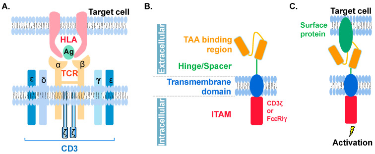
Figure 1.
Schematic view of TCR and CAR molecules. (A) TCR (orange) on T cell is composed of α and β chains and binds to HLA (pink)-Ag (green) complex on target cell. CD3 (blue) has ε, ζ, γ, and δ chains. It serves as a co-receptor of TCR. (B) CAR is composed of the extracellular TAA binding region (orange), hinge or spacer region (green), transmembrane domains (blue), and the intracellular ITAM region (red) which is responsible for activating the cell once TAA binding region senses its target molecules. ITAM can be corresponding domains from CD3ζ or FcεRIγ. Detailed information can be found in Table 1. (C) CAR binds to target protein (green) on cell surface to trigger downstream activation. Noted that, neither HLA nor CD3 is required here.

