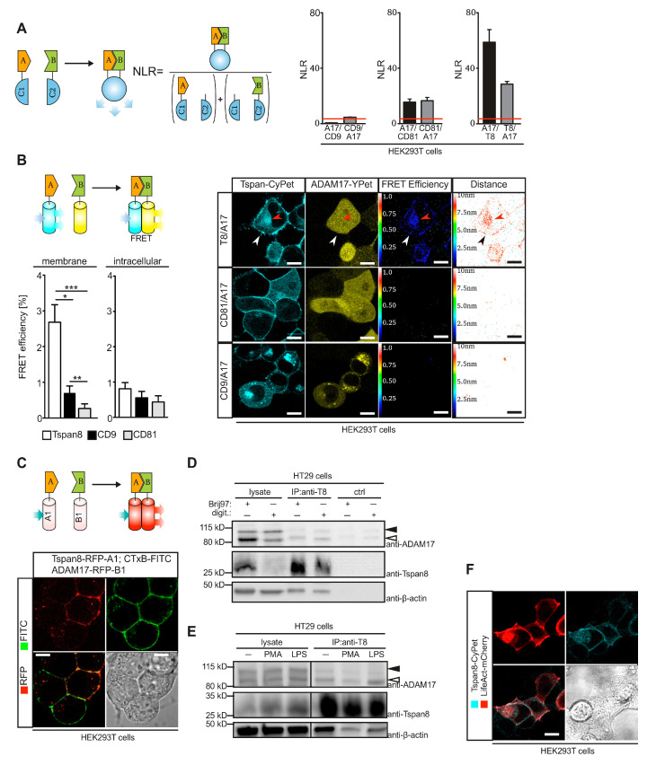
Figure 2.
Tspan8 associates with ADAM17 predominantly at the plasma membrane. (A) Principle of split-luciferase assay. NLR denotes normalized luciferase ratio. ADAM17 specifically interacts with Tspan8 and CD81 but not with CD9 in overexpressing HEK293T cells, as assessed by split-luciferase assay. (B) Interaction of ADAM17 and Tspan8 can be detected at the plasma membrane and in perinuclear compartments using FRET microscopy of HEK293T cells expressing the indicated fusions of ADAM17 or Tspans with the indicated fluorescent proteins. (C) RFP heterodimerization assay confirms ADAM17-Tspan8 interaction at the plasma membrane in transfected HEK293T cells. (D) Endogenous ADAM17 co-immunoprecipitates with endogenous Tspan8 in HT29 colon carcinoma cells. digit.: digitonin (E) Interaction of ADAM17 with Tspan8 is independent of ADAM17 activation. HT29 were stimulated with 1 µg/mL LPS or 200 nM PMA and Tspan8 was isolated by immunoprecipitation and analyzed by SDS-PAGE for co-immunoprecipitating ADAM17. (F) Overexpression of CyPet-fused Tspan8 and mCherry-fused β-actin (LifeAct-mCherry) confirms co-localization of actin filaments and Tspan8 at the plasma membrane. Black filled arrow heads: ADAM17, pro-form, black open arrow heads: ADAM17 mature form. Data are represented as mean ± s.e.m. n = 3 independent experiments (A,B), * p < 0.05, ** p < 0.01, *** p < 0.001, Kruskal–Wallis with Dunn’s post-hoc test (B).

