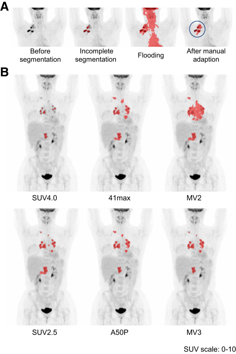
FIGURE 1.
Examples of semiautomatic segmentation. (A) Minimal-intensity projection (MIP) of the PET scan before segmentation; automatic selection with the 41max method missed multiple lesions; adding missing lesions resulted in flooding into the heart, tonsils, and brain; manual adaptation by placing a border around the volume of interest before segmentation resulted in complete selection. (B) Segmentation with SUV4.0 was scored as “missing minor lesions” and “representative delineation.” Segmentation with SUV2.5, 41max, A50P, MV2, and MV3 were scored as “complete segmentation” with “overestimation of delineation.” Segmentation with 41max flooded into the heart and required minor manual adaptation. Segmentation with MV2 flooded into the heart and liver and required major manual adaptations.

