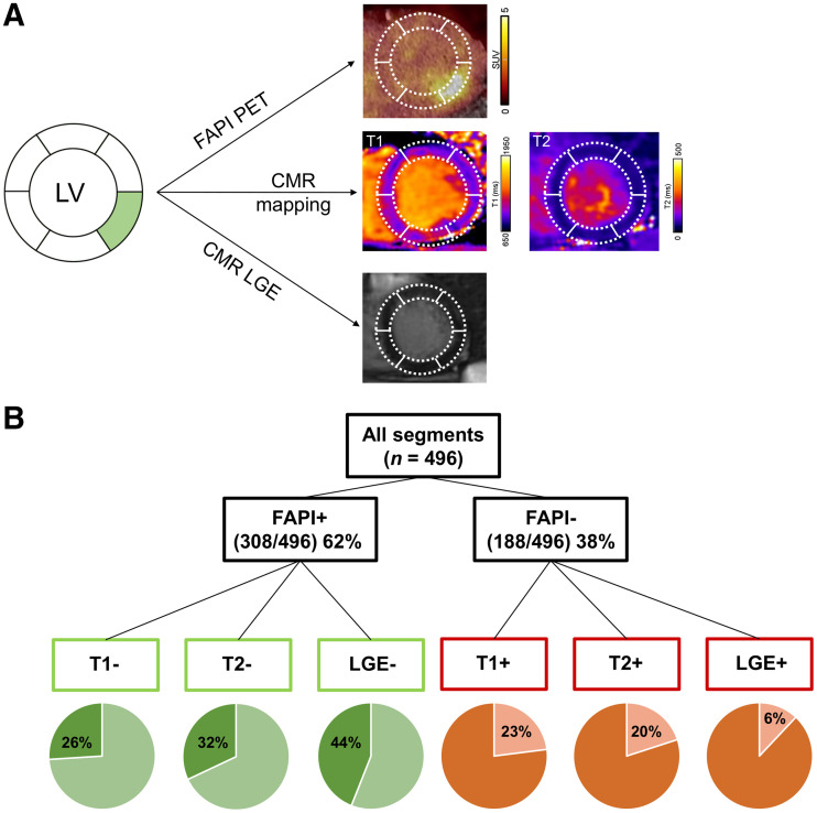
FIGURE 5.
(A) Patient example with FAP-positive, T1-negative, T2-negative, and LGE-negative segment. (B) Segmental comparison of myocardial 68Ga-FAPI-46 distribution, native T1 and T2 mapping, and LGE from CMR, using 16-segment model (apex excluded) in 31 AMI patients with complete datasets, resulting in total of 496 segments.

