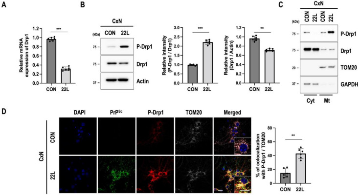
Figure 2.
Scrapie infection induces phosphorylation of Drp1 at Ser616 (P-Drp1), which colocalizes with PrPSc in the mitochondria. (A) The mRNA level of Drp1 was analyzed using qRT-PCR (n = 6, *** p < 0.001; two-sided Student’s t-test). (B) Phosphorylation of Drp1 at Ser616 (P-Drp1) in control (CON) or 22L scrapie-infected (22L) CxN cells at 21 dpi was examined by Western blotting. The intensities of the bands were measured and quantified for each group (n = 6, *** p < 0.001, ** p < 0.01; two-sided Student’s t-test). (C) Drp1 and P-Drp1 levels in the cytosolic (Cyt) and mitochondrial (Mt) fractions. TOM20 and GAPDH were used as markers of the mitochondrial and cytosolic fractions, respectively. (D) The triple-localization of PrPSc (green) with P-Drp1 (red) and TOM20 (white) was determined using confocal microscopy. Quantification of mitochondrial fission by P-Drp1 and TOM20 and mitochondrial translocation was analyzed using EzColocalization plugin for ImageJ software version 1.53a (n = 6, ** p < 0.01; two-sided Student’s t-test). DAPI (blue) was used to counterstain the nuclei; n = number of independent experiments. Enlarged insets (white line boxes) showed the sections at a higher magnification, scale bar, 20 μm. Bullets in bar graph represent datasets from independent experiments.

