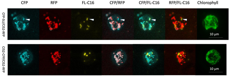
Figure 2.
Co-localization of FL-C16 and CFP-SLI (PTS1 containing peroxisomal targeting protein) was identified in C. reinhardtii. Accumulations of FL-C16 and peroxisomal marker proteins after the cells were cultured in TAP for 24 h in illuminated conditions. Thirty optical sections of each 20 µm z-stack are projected into a plane image with maximum intensity. CFP-SLI: CFP fused to the PTS1 signal peptide serin-leucine-isoleucine. CIS2-RFP: Twenty-five N-terminal amino acid sequences of glyoxylate cycle enzyme citrate synthase 2 (CIS2) fused to RFP. CIS2-CFP: CIS2 fused to CFP. A compartment that contains CIS2-RFP and CFP-SLI but not FL-C16 is shown with a white circle. A pair of compartments that contain FL-C16 and CFP-SLI but not CIS2-RFP are shown with an arrowhead (upper panel). Notice that CIS2-CFP and CIS2-RFP are all colocalized (lower panel), suggesting that non-colocalization between CFP-SLI and CIS2-RFP (marked with the arrowhead) is not an artifact by protein expression or imaging acquisition.

