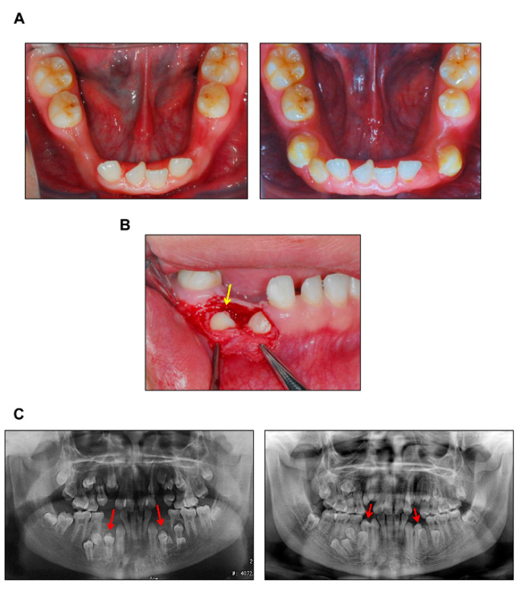
Figure 1.
Guided eruption surgery of CCD patient. (A) Intraoral photographs of CCD patient #5. Left, pre-treatment, 12-year-old; Right, post-treatment, 19-year-old. (B) Surgical exposure of the impacted teeth, 16-year-old. Arrow: right mandibular first premolar. (C) Panoramic radiographs of CCD patient #5. Left, pre-treatment, 12-year-old; Right, post-treatment, 19-year-old. Arrows: mandibular first premolars.

