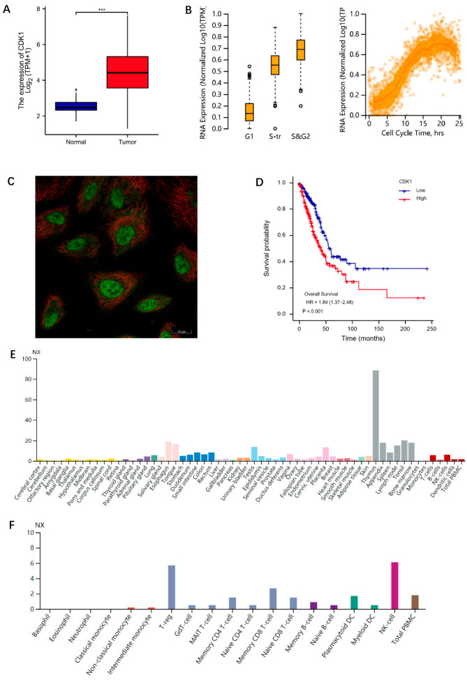
Figure 1.
Information for CDK1. (A) CDK1 expression in normal tissues and LUAD. Significance markers: *** p < 0.001. (B) The expression of CDK1 changed during the cell cycle. (C) The localization of CDK1 in A549 cell. Green for CDK1 antibody; red for tubulin. (D) Correlation between CDK1 expression and OS of LUAD patients. (E) Tissue-specificity analysis. (F) The results of CDK11 expression levels in blood cell samples. The analysis data of (A,D) were from TCGA database. The analysis data of (B,C,E,F) were from HPA database.

