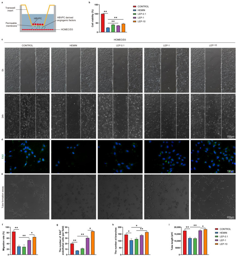
Figure 10.
Pericytes-derived pro-angiogenic factors enhance the survival, migration, proliferation, and tube formation of vascular regeneration following leptin treatment in vitro. (a) Schematic representation of Pericyte and endothelial cell co-culture system. (b) Cell survival was analyzed by CCK-8 assay. (c) Representative images and (f) quantification analyses of HCMEC/D3 migration at 0 h and 24 h after wound scratching. (d) Representative images and (g) quantification analyses of EdU staining in HCMEC/D3. (e) Images of the endothelial tubular networks in Matrigel after 24 h. Analyses of tube formation were done with image J software using the Angiogenesis Analyzer Plugin. Quantification analyses data were shown in (h, i). n = 3 per group. * p < 0.05, ** p < 0.01, between the indicated groups. Error bars, mean ± SEM.

