Figure 2.
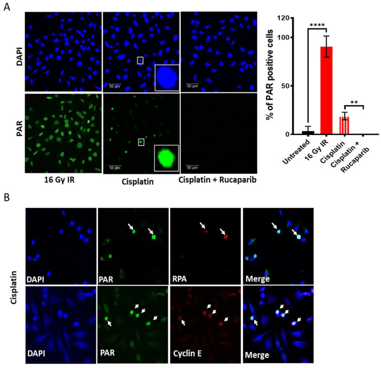
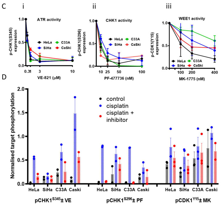
DDR activation by ionising radiation (IR) and cisplatin and inhibition by drugs targeting these pathways: PARP activity was measured by immunofluorescence detection of the product, PAR, in exponentially growing HeLa cells. Cells were fixed immediately after exposure to 16 Gy IR or 6 h incubation with 500 uM cisplatin at 37 °C. (A) Representative images are shown alongside a bar chart of the percentage of PAR positive nuclei, data are mean ± SD from four independent experiments. The difference of PAR positive cells between untreated, IR and cisplatin ± rucaparib treated groups were compared using unpaired t-test. ** and **** are p < 0.01 and 0.0001 respectively. (B) Co-localisation of PAR positive HeLa cells with replication stress marker RPA and cell proliferation marker Cyclin E indicates PARP activation in replicating cells only in response to cisplatin. Images at 40× magnification. (C) pCHK1S345, pCHK1S296, and pCDK1Y15 levels in cells treated with increasing concentrations of (i) VE-821, (ii) PF-477736 and (iii) MK-1775, respectively (Supplementary Figure S4). Data represents mean ± SEM (N = 3). (D) Analysis of ATR, CHK1 and WEE1 activity from densitometric analysis of pCHK1S345, pCHK1S296, and pCDK1Y15, respectively, in Western blots of exponentially growing cell exposed to 0.5% DMSO alone, 3 μM cisplatin and 3 μM cisplatin + 1 μM VE-821 (VE), 50 nM PF-477736 (PF) or 100 nM MK-1775 (MK) for 24 h prior to harvest cells and lysate preparation (Supplementary Figure S4). Data represents mean of two independent experiments.


