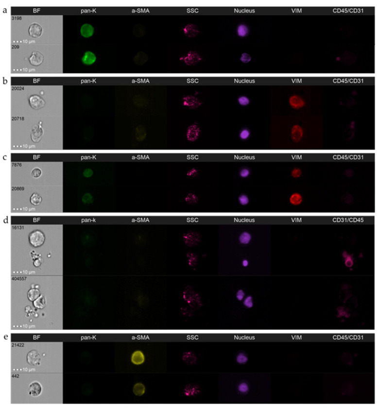
Figure 1.
Representative pictures of the spectrum of epithelial-to-mesenchymal phenotypes in circulating tumor cells are detected using imaging flow cytometry in breast cancer patients: (a) epithelial CTC, (b) mesenchymal CTC, (c) epithelial–mesenchymal CTC, (d) double negative CTC, and (e) circulating cancer-associated fibroblasts. BF—brightfield; pan-K—pan-keratins; a-SMA—alpha-smooth muscle actin; SSC—side scatter; Vim—vimentin; CD45/CD31—leukocyte/endothelial cell marker. Amnis® ImageStream®X Mk II (Luminex, Austin, TX, USA), objective 40×.

