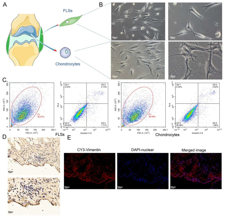
Figure 4.
Labeling of FLSs in SM, and cell viability detection for isolated FLSs and chondrocytes. (A) FLSs and chondrocytes obtained from SM and AC of rats, respectively. (B) Observation of FLSs and chondrocytes using light microscopy. (C) FCM was used to determine the viability of FLSs and chondrocytes. (D) Vimentin IHC staining of SM. (E) Vimentin IF staining of SM.

