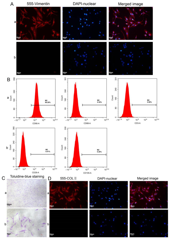
Figure 5.
Characterization of the synoviocytes and chondrocytes. (A) Vimentin IF staining of FLSs: (a) test cells; (b) isotype controls. (B) The surface markers were analyzed using FCM: (a) the synoviocyte surface markers of CD90 (FLSs), CD68 (macrophages), and CD3 (T cells); (b) the MSC surface markers of CD29 and CD105. (C) Characterization of the chondrocytes using toluidine blue staining. (D) Characterization of the chondrocytes with COLII immunofluorescence analysis: (a) test cells; (b) isotype controls.

