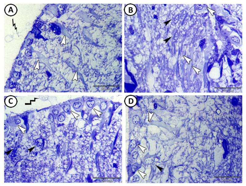
Figure 2.
Semithin sections of the ependymal cells (ECs) were stained with toluidine blue. (A) The processes of ECs (white arrowheads) extended through the full thickness of the tectal laminae. Note the apical neurosecretion (zigzag black line). (B,C) The basal processes of the EC (white arrowheads) were connected with stem cells (black arrowheads). Note that the surfaces of the ECs were covered with secretion (zigzag black line). (D) The interaction between the ECs (white arrowheads) and multipolar neurons (black arrowheads).

