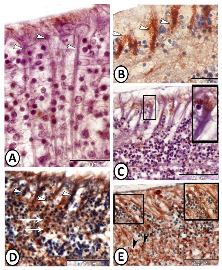
Figure 4.
Immunohistochemistry of ECs. (A) ECs showed expression of IL-1β (white arrowheads) in their cytoplasmic processes. (B) ECs showed strong cytoplasmic expression of APG-5 (white arrowheads). (C) ECs showed nuclear expression of Nfr2 (boxed areas). (D,E) ECs showed expression of myostatin (white arrowheads) and SOX-9 (boxed areas), respectively, in their cytoplasmic processes. Note the proliferative activity of the neighboring stem cells (black arrowheads and white arrows).

