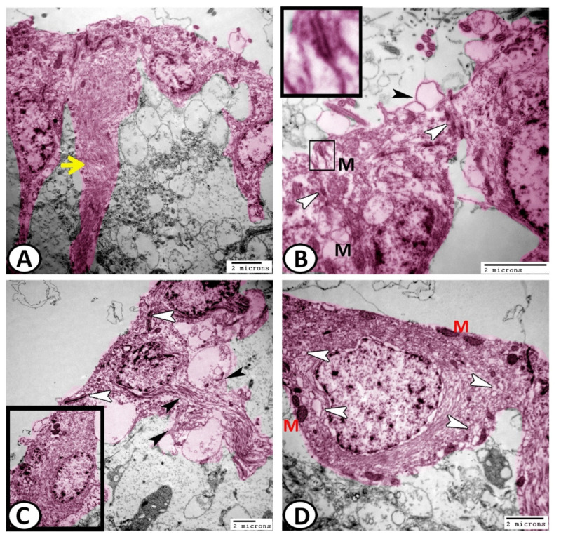
Figure 7.
Digital colored transmission electron microscopy (TEM) of ECs (pink). (A) The ECs showed bundles of intermediate filaments (yellow arrowhead) in their processes and basal poles. (B) The ECs were connected by desmosomes (boxed areas) and gap junctions (white arrowheads). Note the presence of surface membrane-bounded vesicles (black arrowhead) containing neurosecretion and mitochondria (M). (C) The ECs showed distinct fascicle filaments that contained intermediate filaments (black arrowheads). Note the junctional complexes between the cells (white arrowheads). (D) Higher magnification of the boxed area of (C) shows the non-ciliated ECs that possessed many mitochondria (M) and abundant vesicles (white arrowheads).

