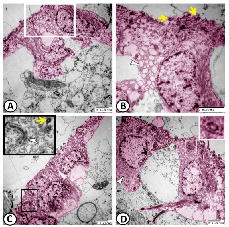
Figure 8.
Digital colored transmission electron microscopy (TEM) of ECs (pink). (A,B) The surfaces of ciliated ECs showed many cross-sections of cilia (yellow arrowheads). The abluminal and lateral cell surfaces of ECs showed pinocytotic activities with many coated vesicles (white arrowhead). (C) The apical cytoplasm of ECs possessed centriole (yellow arrowhead) and well-developed junctional complexes (white arrowhead in the boxed area). (D) The cytoplasm of these ECs also contained bundles of intermediate filaments (white arrowhead), distinct fascicle filaments (black arrowhead) and centrioles (boxed areas).

