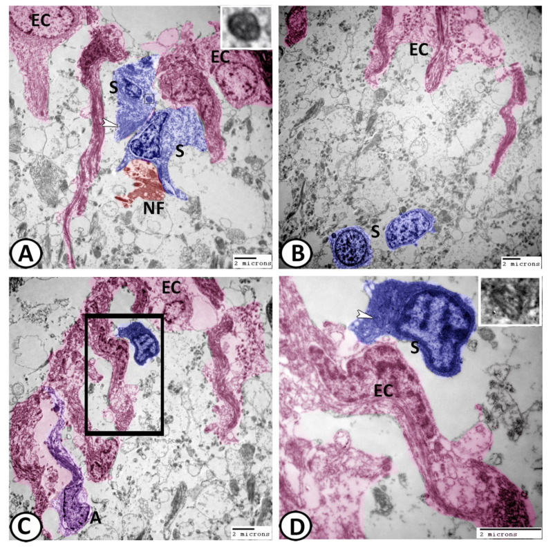
Figure 9.
Digital colored transmission electron microscopy (TEM) of ECs (pink), astrocytes (violet), and stem cells (blue). (A,B) Many stem cells (blue, S) could be demonstrated among the ECs. In (A), bundles of intermediate filaments (white arrowhead) and centrioles (boxed areas) were observed in the cytoplasm of stem cells, while generated axons (NF, red) were observed in direct contact with these stem cells. (C) The processes of astrocytes (A, violet) and stem cells (blue) were seen among the ECs. (D) In addition, the stem cell cytoplasm contained centrioles (boxed area, white arrowheads).

