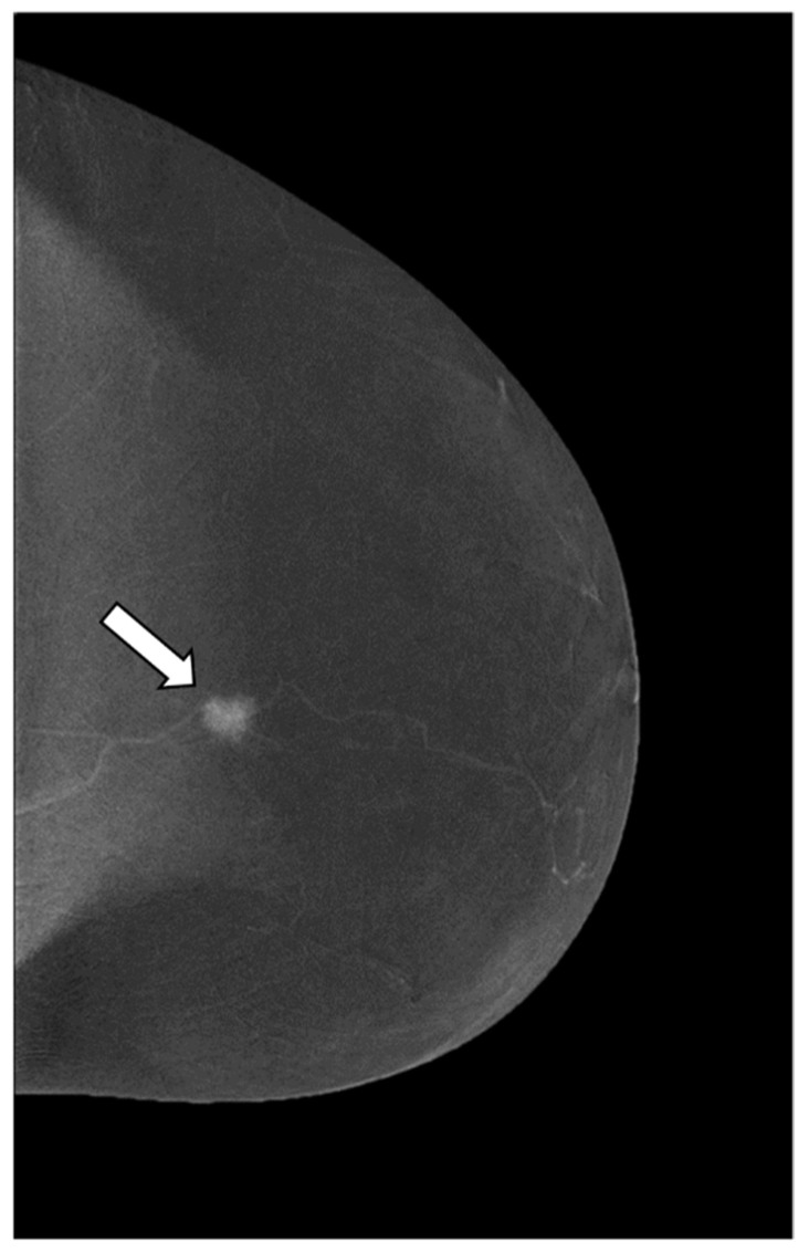
Figure 2.
Craniocaudal (CC) view of CESM subtraction image of a 59-year-old patient with a suspicious lesion of the left breast measuring less than 10 mm (BI RADS 4c). The subtraction images show a mass enhancement marked, irregular, heterogeneous, and unpurified (enhancement score 4) (white arrow). The histological result is an invasive ductal carcinoma.

